Friday, December 19, 2014

3D-printed tumor replicas to better measure doses of cancer-fighting drugs

December 18, 2014
3D printing models of tumors and their surrounding organs could help specialists to delive...
3D printing models of tumors and their surrounding organs could help specialists to deliver cancer-fighting drugs with greater accuracy (Photo:Shutterstock)
Administering the correct dosages to fight cancerous tumors can be a difficult balancing act. Too much of the radioactive drugs can cause harm to healthy tissue, but not enough will see the cancer cells survive and continue to spread. But a new technique developed at The Institute of Cancer Research in London may afford doctors an unprecedented level of accuracy in performing radiotherapy, using 3D-printed replicas of a patient’s organs and tumors to better determine how much radiation a tumor has received.
In preparing for radiotherapy, using models of a tumor is not uncommon, but these are typically hand-made and aren’t always so reliable.
"The big challenge we faced was to produce a model that was both anatomically accurate and allowed us to monitor the dose of radiation it received," says Dr Jonathan Gear, Clinical Scientist at The Institute of Cancer Research. "We found that the printed replicas could give us information we couldn’t get from 2D scans, you will always get more information from a 3D model than a flat image."
Gear and his fellow researchers are physicists working in molecular radiotherapy, a method used in treating thyroid cancer, adult neuroendocrine tumors, childhood neuroblastoma and bone metastases from prostate cancer. Using scans taken during patient treatment, the team 3D printed replicas of a tumor and surrounding organs. Dubbed "phantoms," the plastic models were filled with the same radioactive liquid given to patients and then monitored to see the likely effects of radiotherapy in that particular patient.
The researchers say that in their initial testing, the method allowed them to more precisely calculate the dose of radiation received by the patient and then adjust the following treatments accordingly. While promising, the scientists say that further testing in larger studies is needed, but if successful, the technique could greatly improve the precision of molecular radiotherapy.
"We’ve seen reports on how 3D printing is being used for prosthetics and to inform surgery, and this research shows it has the potential to improve cancer treatment too, by helping us to perform complex radiotherapy calculations more accurately," says Dr Glenn Flux, Head of Radioisotope Physics at the Joint Department of Physics at The Institute of Cancer Research. "We’re really excited by this technology and the potential it has for personalizing cancer treatment with highly targeted radiation."
This ability to customize treatments extends well beyond cancer therapies. Earlier this year we saw a 3D printed replica of an baby’s defected heart help surgeons prepare for what would be a life-changing surgery. It has also allowed surgeons to develop customized medical implants, including everything from a titanium heel to an upper jaw prosthesis.

Thursday, December 18, 2014

FDA Approves Gardasil 9: More is Better for Cervical Cancer Prevention

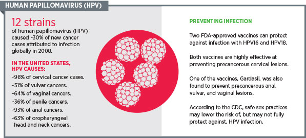
Last week, the U.S. Food and Drug Administration (FDA) announced the approval of a new vaccine—Gardasil 9—that prevents infection with five more cancer-causing subtypes of human papillomavirus (HPV) than the current HPV vaccines, Gardasil and Cervarix.
As discussed on this blog by my AACR colleague Srivani Ravoori, PhD, research shows that Gardasil 9 could prevent almost 90 percent of cervical cancer cases worldwide. It also has the potential to prevent more cases of vulvar, vaginal, and anal cancers than Gardasil and Cervarix can.
The reason Gardasil 9 has the capability to prevent more cases of cancer than Gardasil and Cervarix is that while Gardasil and Cervarix prevent infection with the two main cancer-causing HPV subtypes, HPV16 and -18, Gardasil 9 protects against an additional five cancer-causing HPV subtypes: HPV31, -33, -45, -52 and -58. Gardasil and Gardasil 9 also prevent infection by two subtypes of HPV that cause genital warts, HPV6 and -11.
Gardasil 9 is approved for the prevention of cervical, vulvar, vaginal, and anal cancers caused by HPV16, -18, -31, -33, -45, -52, and -58. It is likely that Gardasil 9 could also prevent a significant number of cases of cancers of the oropharynx—the part of the throat just behind the mouth—because more than 50 percent of U.S. cases of these cancers are related to HPV infections. However, research is needed to show that this is indeed the case.
1409119_AACRblog_HPV_3
Unfortunately, Gardasil 9 will only live up to its promise if those individuals for whom it is recommended—females ages 9–26 and males ages 9–15—get vaccinated. With the latest data from the Centers for Disease Control and Prevention estimating that just 38 percent of adolescent girls (ages 13–17) and 14 percent of adolescent boys completed a course of Gardasil or Cervarix in 2013, it is clear that more needs to be done to develop and implement effective strategies.
A number of recommendations to increase HPV vaccine update were made in the most recent President’s Cancer Panel Report, “Accelerating HPV Vaccine Uptake: Urgency for Action to Prevent Cancer,” which was released in February 2014, and there is optimism that we can make progress.
“We can prevent a generation of young Americans from having to go through the same experience I did,” said Bob Margolis, a seven-year survivor of HPV-related cancer and advocate for HPV awareness and vaccination, in the AACR Cancer Progress Report 2014.
 Learn more about Margolis’ story in this video.

Kicking Cancer with Technology


Kicking Cancer with Technology
While we know that some behaviors like smoking cigarettes, neglecting to wear sunblock, and eating chemically processed foods can increase our chances of cancer, there is no sure-fire way of preventing the disease from attacking our bodies. A nonsmoker isn't immune to lung cancer, just as a person who never stepped foot in a tanning salon may still be diagnosed with melanoma. It's a game of chance, and the innovators at the German National Center for Tumor Diseases (NCT) and SAP aren't willing to leave this battle up to fate.
By 2035, it's estimated that 22 million people will have cancer and the disease will account for 13 million deaths worldwide (Source:World Health Organization). In addition, the cost of developing treatment is rising, the number of new cancer drugs is declining, and the drugs currently available are only effective in about one fourth of treatments.
Why is cancer so deadly and difficult to cure? Because almost every instance of cancer is unique, and each individual occurrence responds differently to treatment. There is no exact way of knowing which treatments will work for a specific patient. Therapies are administered to patients based on a doctor's past experience and by manually reviewing, parsing, and consolidating the relevant information, typically in spreadsheets. Such manual processes can cause significant delays for patients waiting for care, and for a cancer patient, time is vital; they can't afford to wait.
NCT's goal is to administer specialized treatment suited to a patient's specific molecular profile as quickly as possible. NCT focuses on cancer research as well as patient treatment. In just one year, NCT touches:
  • 10,000 patients
  • 15,000 courses of treatment
  • 50,000 appointments
This means that a single patient can have up to 1,200 pieces of data in their record. In total, NCT must manage up to 150,000 data sets and 3.6 million data points, a process that, if done manually, could take weeks. However, the analysis can be done in real time using the center's Patient Data Explorer tool supported by the SAP HANA platform. NCT is significantly accelerating the development of individual, highly adjusted cancer therapies.
Beating Breast Cancer: Listen to how NCT is helping one woman win the battle over breast cancer by providing her the right medication to fight her disease.
With Patient Data Explorer, patient data from different sources such as clinical information systems, tumor registries, biobank systems, and even text documents (like physicians' notes) can be accessed, analyzed, and put into actionable context in minutes rather than days. In addition, the tool offers a comprehensive overview of each individual patient's medical history in a graphical timeline, making it easy to access information with any level of detail.
"We are now able to extract and analyze data from various sources and present it to physicians and researchers in a way that enables surprising new insights," says Professor Dr. Christof von Kalle, Director of the Department of Translational Oncology, as he explains the importance of continued research and collaboration with SAP HANA to analyze huge amounts of data in real time. "In the future, we would like to use the SAP HANA platform for every diagnostic and therapeutic step, because every cancer is different and can vary immensely from one patient to the next."
Unfortunately cancer isn't going to miraculously disappear. But with leading innovators like NCT teaming with SAP, we are creating technologies that will help find new treatments and determine the most effective existing ones. By quickly providing patients with specialized treatment, we are combating this ugly disease harder than ever. Knowing that such in-depth scientific progress has been made and continues, I have much confidence that we will someday be able to save all the moms, dads, grandparents, children, and babies in the world who are diagnosed with cancer.
Read the original story on SAP Business Trends.

Friday, December 12, 2014

The Punishing Cost of Cancer Care

Photo
CreditStuart Bradford
“I think it’s time to switch therapies,” I told my patient, as he and his wife sat next to each other by the wall of my exam room.
He stared at me, unblinking, through his chipped, wire-rimmed glasses as his wife looked quickly down at the medication list she clutched in her hands. Her worn purse sat on the floor by her chair. “Switching therapies” was a euphemism for “your cancer is progressing,” a point I didn’t need to hammer home with them – they both knew the score.
“O.K.” he said, taking a deep breath. “What’s next?”
We had been navigating his cancer together for a year-and-a-half, balancing the most effective treatments we could devise with what his health insurance would cover, and what he could afford. The 90-mile drive to Cleveland was itself an economic strain, gas prices being what they were and their aging car becoming increasingly more finicky, particularly during the cold winter months.
“The next drug I’d suggest is a pill that you’d take every day. It can get you into a remission, and may even allow you to live a little longer. It’s the one we’ve talked about before,” I told him. He nodded, remembering.
“The real expensive drug?” he asked.
This time I nodded. When I first suggested he take this pill as part of his chemotherapy regimen, I warned him about the possible cost, as this has become a part of my standard informed consent process. I review potential risks, benefits, alternative medications, the people involved in a patient’s care, and now economic risks.
After checking with his insurance plan, which did not cover chemotherapy pills the way it did intravenous chemotherapy, I had the unpleasant task of telling my patient that the medication would cost him $5,000 per month. At the time he had laughed, a mixture of surprise and incredulity. He was too well-off to qualify for patient assistance, but too poor to afford the drug. His face was grave now.
“I won’t bankrupt my family for a month or two more time,” he said. “I have to leave them something.” His wife looked up briefly, about to interject, but remained quiet. This was ground they had already covered, and she would make her case for his taking the pill when I wasn’t present. They were private people, proud, and wouldn’t disagree with each other in front of someone who wasn’t family.
As the price of chemotherapy now routinely reaches $100,000 for a full treatment course, my patients are forced more and more into making the equivalent of Sophie’s Choice when it comes to treating their cancer: Spend down their savings for an improvement in survival that might amount to a few weeks, secretly hoping that they will be one of the lucky few at the “tail” of the survival curve – the handful of people who live years more; or decline the therapy and in so doing ensure that their families will be provided for after they have died.
I had to wonder whether anyone should be forced to make such a decision.
My patient and I discussed a couple of other treatment options, and ways that he might receive his therapy closer to home, a plan that made him visibly relax.
“Not that we don’t like seeing you,” he joked. His wife didn’t smile, though. Perhaps she wasn’t ready yet to let him go, or didn’t agree with his reasoning that she would be destitute if he chose the pill. Maybe she was incensed that hard-working people like themselves would be placed in such a quandary precisely at the time of their retirement, and in such a wealthy nation. The way she set her mouth, I guessed it was a combination of all three.
“I’ll miss seeing you,” I told them both as we all got up to leave. There was nothing else I could say – this was the part of cancer care I didn’t have the tools to fix.
http://well.blogs.nytimes.com/2014/12/11/the-punishing-cost-of-cancer-care/?emc=edit_tnt_20141212&nlid=52389906&tntemail0=y&_r=0

Tuesday, December 9, 2014

Scientists make breakthrough in understanding of rare blood cancer

Scientists make breakthrough in understanding of rare blood cancer
New breakthrough in splenic marginal zone lymphoma (SMZL)
A breakthrough in understanding of a rare form of blood cancer could lead to significant improvements in treatment for patients. Scientists from the University of Southampton announced their findings at a prestigious scientific conference in San Fransisco on Sunday 7 December.
The team used various sophisticated 'gene sequencing' techniques to screen DNA from 175 patients with splenic marginal zone (SMZL), a  of the immune system. They identified key genetic errors that can influence the speed at which the cancer develops in different patients.
SMZL generally develops slowly. After diagnosis patients are monitored and only start treatment when the disease reaches a stage when it is needed. It is a , accounting for only 3% of all cases of lymphoma, and there has been little research into the disease until now.
Professor Jonathan Strefford presented his team's findings at the American Society of Hematology's Annual Meeting. The research was funded by the  charity Leukaemia & Lymphoma Research.
Professor Strefford, of the University of Southampton, said: "By revealing the genetic signs linked to a poor prognosis, doctors will be able to determine which patients have more aggressive cancer and require higher doses of drugs or treatment at an earlier stage."
The scientists identified, for the first time, how individual gene faults connected to SMZL can influence a patient's prognosis. Errors in the TP53 gene in  were shown to be 'drivers' of the cancer, potentially acting as a warning for doctors of shorter survival times. The presence of faults in the KLF2 and NOTCH2 genes indicated that patients generally required intensive treatment for their disease more quickly.
Dr Matt Kaiser, Head of Research at Leukaemia & Lymphoma Research, said: "Understanding how different genetic changes contribute to a patients' disease has allowed us to make huge strides in designing treatments for many different blood cancers, but until now splenic marginal zone lymphoma has been a bit behind. This research could help doctors and patients make more informed choices about treatment plans, and help to significantly improve the outlook for this group of ."

Toughest breast cancer may have met its match

breast cancer
Mammograms showing a normal breast (left) and a cancerous breast (right). Credit: Wikipedia.
Triple-negative breast cancer is as bad as it sounds. The cells that form these tumors lack three proteins that would make the cancer respond to powerful, customized treatments. Instead, doctors are left with treating these patients with traditional chemotherapy drugs that only show long-term effectiveness in 20 percent of women with triple-negative breast cancer. Now, researchers at The Johns Hopkins University have discovered a way that breast cancer cells are able to resist the effects of chemotherapy—and they have found a way to reverse that process.
A report of their findings was published online in the journal Proceedings of the National Academy of Sciences on Dec. 1.
Triple-negative breast cancers account for about 20 percent of all breast cancers in the United States, and 30 percent of all breast cancers in African-American women. In addition to being resistant to , they are known to include a high number of , which are responsible for relapses and for producing the metastatic tumors that lead to the death of patients with cancer. Previous research revealed that triple-negative breast  show a marked increase in the activity of many genes known to be controlled by the protein hypoxia-inducible factor (HIF). Given these past results, a research team directed by Gregg Semenza, M.D., Ph.D., decided to test whether HIF inhibitors could improve the effectiveness of chemotherapy.
"Our study showed that chemotherapy turns on HIF and that HIF enhances the survival of breast cancer stem cells, which are the cancer cells that must be killed to prevent relapse and metastasis," says Semenza, the C. Michael Armstrong Professor of Medicine at Johns Hopkins and a Johns Hopkins Kimmel Cancer Center expert. "The good news is that we have drugs that block HIF from acting."
Semenza's study began by treating lab-grown triple-negative  with the chemotherapy drug paclitaxel and looking for changes in HIF levels. After four days of treatment, HIF protein and activity levels had increased, as had the percentage of breast cancer stem cells among the surviving cells. When Semenza's team, led by postdoctoral fellow Debangshu Samanta, Ph.D., genetically altered the cancer cells to have less HIF, the cancer stem cells were no longer protected from death by chemotherapy, demonstrating that HIF was required for the cancer stem cells to resist the toxic effects of paclitaxel, Semenza says.
At the molecular level, the team found that one of the ways HIF enhances the survival of the stem cells is by increasing the levels of a protein, multidrug resistance protein 1 (MDR1), which acts like a pump to expel chemotherapy from cancer cells. However, when triple-negative breast cancer cells were given paclitaxel plus the HIF inhibitor digoxin, MDR1 levels went down rather than up.
In mice that were implanted with triple-negative breast cancer cells, treatment with digoxin and paclitaxel decreased tumor size by 30 percent more than treatment with paclitaxel alone. The combination therapy also decreased the number of breast  and the levels of MDR1. Treatment with digoxin plus a different chemotherapy drug, gemcitabine, brought tumor volumes to zero within three weeks and prevented the immediate relapse at the end of treatment that was seen in mice treated with gemcitabine alone.
Analysis of patient databases showed that among women with  who are treated with chemotherapy, those with higher-than-average levels of HIF activity in their tumor were much more likely to die of breast cancer than those with lower-than-average HIF levels. Samanta notes that the HIF inhibitor digoxin is already approved by the Food and Drug Administration for treating heart failure. Several other drugs that inhibit HIF have also been identified and are currently being tested in patients with cancer. If the team's work is verified in clinical trials, the researchers think that potentially unresponsive patients could be identified before treatment and given a more effective combination therapy.

Saturday, December 6, 2014

‘True risk’ of breast cancer: MRI precisely pinpoints chances of developing disease

HUTCH NEWS

‘True risk’ of breast cancer: MRI precisely pinpoints chances of developing disease

Study finds a screening byproduct, once thought to be clinically insignificant, is useful in predicting the likelihood of breast cancer in high-risk women

Dec. 4, 2014
Breast MRI
A feature of breast imaging via MRI, termed “background parenchymal enhancement,” or BPE, can help pinpoint the chances of developing breast cancer in high-risk women. Left: An MRI image of a woman with high BPE (shown as scattered white dots throughout her breasts). This patient developed breast cancer three years after her exam. Right: An MRI image of a woman with low BPE, who has not developed breast cancer in the five years since this image was taken.
Image courtesy of the University of Washington Breast Imaging program
A new study, using a built-in but little-understood visual byproduct of MRI breast imaging, offers the possibility of nailing down a woman’s “true risk” of breast cancer – for a woman already at high risk of the disease, that is.
One in eight U.S. women will develop breast cancer over her lifetime, according to theNational Cancer Institute, meaning the average woman has about a 12 percent chance of getting the disease. Women termed “high risk” are those with a 20 percent or greater lifetime chance of breast cancer.
But most women, and their doctors, are left without much information beyond that high- versus normal-risk status, said Dr. Habib Rahbar, a radiologist at Seattle Cancer Care Alliance, Fred Hutchinson Cancer Research Center’s treatment arm, and one of the study’s investigators. It turns out being dubbed high risk includes a wide range of probabilities of actually developing the disease.
“Twenty percent to 100 percent is obviously quite a great range, so the ability to further determine which patients are most likely to develop breast cancer is really significant,” Rahbar said.
Knowing where a woman falls within that wide range would help her doctors better guide her screening and treatment options, he said.
Rahbar and his colleagues found that a feature of breast MRI imaging that previously was not known to have clinical implications can in fact help pinpoint the chances of developing breast cancer among those at high risk. That tool could lead to a more precise cancer-risk assessment. Dr. Brian Dontchos, a Seattle radiologist and study investigator, presented the findings today at the Radiological Society of North America’s annual meeting in Chicago.

MRI might predict cancer before it starts

Their study suggests that breast MRI, a technique commonly used for screening high-risk women, could be used not only to catch breast cancer, but to predict it before it starts.
To look for breast cancer, radiologists compare MRI images of the breast before and after injecting a contrast agent into the patient’s bloodstream; tumors glow bright when the dye pumps through the breast’s blood vessels. But it turns out for some women, normal parts of their breasts light up too. That background glow, which radiologists term “background parenchymal enhancement,” or BPE, has largely been ignored except as an inherent artifact of some MRIs.
“No one really understood whether BPE had any clinical implications,” Rahbar said.
The researchers tracked 487 high-risk women who received screening MRIs at SCCA. Over the six years of the study, 23 of those women developed breast cancer. The researchers compared those women with an age-matched group of cancer-free women from the study and found that those who went on to develop cancer had higher BPE levels on their initial MRI exam than those who didn’t develop breast cancer.
The researchers don’t yet know whether BPE could also predict breast cancer risk in the general population. They hope to examine that question in future research, but to do so would be a much larger and more costly undertaking, Rahbar said, in large part because insurance companies don’t currently cover the cost of MRI screening for average-risk women.
“This opens up an avenue in imaging that hasn’t been well explored,” added Dr. Savannah Partridge, a co-investigator of the study and breast imaging expert at SCCA and UW Medicine.

A powerful marker of cancer risk

Current methods to calculate breast cancer risk are based on studies of populations, Rahbar said. That’s a problem when it comes to assessing an individual woman’s risk or making medical decisions based on that risk.
Doctors use a woman’s age, family history and facts about her menstrual cycle and whether she’s had children (both of which give clues as to how much estrogen has been kicking around in her body) to determine breast cancer risk.
“The problem with those factors is that they’re really common in the general population, and they’re individually very weak predictors of whether a woman will develop breast cancer or not,” Rahbar said. “[BPE] could be a very powerful individual marker.”
If doctors can uncover a woman’s true risk of breast cancer, they can help her make concrete decisions about managing that risk. Since nearly all radiologists experienced in breast MRI understand how to identify BPE when performing breast MRIs, any facility that performs screening MRIs could potentially use this tool, Rahbar said.
Depending on how great a woman’s chances of developing the disease, her options would range from increased screening to taking a daily dose of a cancer-preventing drug, such as tamoxifen, to a preventive double mastectomy. Some of those choices would be more appropriate for women at very high risk of cancer but possibly not for those just over the borderline of high risk, Rahbar said.
“We don’t want to provide those sorts of recommendations to patients without having a really clear idea what their risk is rather than this huge range,” he said.

A fertile ground for cancer growth

What BPE is, and why it could herald cancer’s arrival, remains a bit mysterious. Since it’s related to blood flow, scientists feel it reflects some kind of activity in the breast.
BPE is also loosely related to breast density, another known risk factor for breast cancer. Breast density refers to how much of a woman’s breasts are made up of fat versus glandular and connective tissue. The less fat, the higher the density. A woman with very low density is unlikely to show any of this background signal on an MRI, Rahbar said, but not all women with dense breasts will have high BPE.
The researchers also found that among high-risk women, breast density was an unreliable predictor of breast cancer risk and that BPE may prove to be a better tool for measuring such a woman’s chances of getting breast cancer.
As for why BPE correlates with cancer risk, that’s unknown, but the researchers hypothesize that it’s related to the pro-cancer effect of certain circulating hormones. .In pre-menopausal women, the MRI signal rises and falls with the ebbs and flows of estrogen in the menstrual cycle. Women with lower estrogen levels – post-menopausal women and those taking the hormone suppressor tamoxifen – have low BPE.
Because lifetime estrogen exposure is itself a risk factor for breast cancer, Rahbar thinks high BPE might be a way to visualize an estrogen-sensitive – and thus more cancer-prone – breast.
“In many ways, we feel like we may be looking at areas within breasts that are very fertile for a cancer to grow,” he said.
The International Society for Magnetic Resonance in Medicine and the Roger E. Moe Fellowship in Multidisciplinary Breast Cancer Care funded the research.
Editor’s note: SCCA is recruiting high-risk women with dense breasts for a clinical trial that aims to improve breast cancer detection among this group.
Dr. Rachel Tompa, a staff writer at Fred Hutchinson Cancer Research Center, joined Fred Hutch in 2009 as an editor working with infectious disease researchers and has since written about topics ranging from nanotechnology to global health. She has a Ph.D. in molecular biology from the University of California, San Francisco and a certificate in science writing from the University of California, Santa Cruz. Reach her at rtompa@fredhutch.org.
Are you interested in reprinting or republishing this story? Be our guest! We want to help connect people with the information they need. We just ask that you link back to the original article, preserve the author’s byline and refrain from making edits that alter the original context. Questions? Email senior writer/editor Linda Dahlstrom at ldahlstr@fredhutch.org.
Solid tumors, such as those of the breast, are the focus of Solid Tumor Translational Research, a network comprised of Fred Hutchinson Cancer Research Center, UW Medicine and Seattle Cancer Care Alliance. STTR is bridging laboratory sciences and patient care to provide the most precise treatment options for patients with solid tumor cancers.

Monday, December 1, 2014

Hair Loss and Medical Treatment


“I have to tell you I love my new hair and people come up to me and want to know where I have my hair done!” Susan’s Special Needs client thank you note.

“Finding a wig at a difficult time in your life should be a comforting and secure experience.” explains Susan Thomas Oncology Nurse Clinician and Breast Cancer Survivor.

It’s not just a wig at Susan’s. It’s about a team approach helping our clients feel safe in privacy, welcomed and comforted when they walk in the door. Our store offers unique wigs not found “just anywhere”. All of our wigs carry a special brand of Susan’s excellence and quality. Our goal is to educate our clients presenting them with options and help them determine what is best for them, their lifestyle and their budget.

A wig is not just a wig – it’s the talent of our wig stylists, Verna Haak, Karen Ashmore and Shannon Forgash licensed cosmetologist who share Susan’s passion to serve and provide the expertise of fitting and styling the wig to customize each wig for each client-included in price of wig. Collectively they offer 34 years of experience in our unique wig custom tailoring process. Our wig fitting appointments are approximately 1 ½ hours helping to ensure a perfect fit. The client’s scalp and wig are evaluated for comfortable fit, wig length, density and volume – all to meet our client’s expectations. Our stylists review wig care instructions and provide tips for keeping their new wig healthy.

You will also find a stylish selection of headwear including hats, turbans, sleep caps and scarves presenting contemporary and fashionable choices. Visit our New York Fashion Bazaar.


“What happens here is pretty remarkable,” says Susan. “We are thankful to be able to share in this important aspect of our client’s journey.”